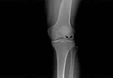
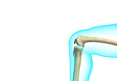
Knee Arthritis
The joint surface is covered by a smooth articular surface that allows pain-free movement in the joint. Arthritis is a general term covering numerous conditions where the joint surface or cartilage wears out. This surface can wear out for several reasons; often the definite cause is not known. Arthritis often affects the knee joint. When the articular cartilage wears out, the bone ends rub on one another and cause pain. The most common type of arthritis is osteoarthritis. It occurs with aging and use.
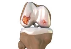
Knee Osteoarthritis
Osteoarthritis also called degenerative joint disease, is the most common form of arthritis. It occurs most often in older people. This disease affects the tissue covering the ends of bones in a joint (cartilage).In a person with osteoarthritis, the cartilage becomes damaged and worn out causing pain, swelling, stiffness and restricted movement in the affected joint. This condition most commonly affects the joints in the hips, knees, hands, and spine. Rarely, the disease may affect the shoulders, wrists, and feet.
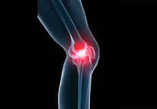
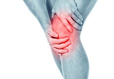
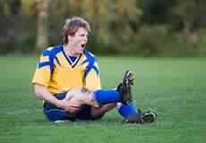
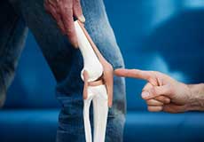
Knee Pain
Knee pain is a common condition affecting individuals of various age groups. It not only affects movement but also impacts your quality of life. An injury or disease of the knee joint or any structure surrounding the knee can result in knee pain. A precise diagnosis of the underlying cause is important to develop an appropriate treatment plan.
Knee Injury
Pain, swelling, and stiffness are the common symptoms of any damage or injury to the knee. If care is not taken during the initial phases of injury, it may lead to joint damage, which may end up destroying your knee.
Knee Sprain
Knee sprain is a common injury that occurs from overstretching of the ligaments that support the knee joint. A knee sprain occurs when the knee ligaments are twisted or turned beyond its normal range, causing the ligaments to tear.
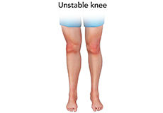
Unstable Knee
Damage to any of these supportive structures causes instability of the knee joint. An unstable knee can be caused by the sudden twisting of the knee, tears of the meniscus, ligament or capsule, osteoarthritis of the knee (wear and tear of the cushioning cartilage tissue between the bones) and sports injuries. When these tissues get injured, the patella or kneecap can move out of its groove in the knee joint and lead to instability.
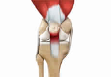
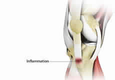
Jumper's Knee
Jumper’s knee, also known as patellar tendinitis, is inflammation of the patellar tendon that connects your kneecap (patella) to your shinbone. This tendon helps in the extension of the lower leg.
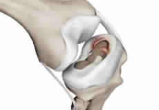
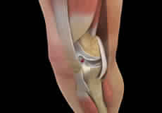
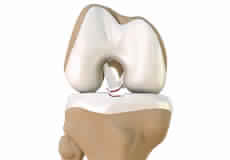
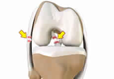
Meniscal Tears
A meniscal tear is a common knee injury in athletes, especially those involved in contact sports. A sudden bend or twist in your knee causes the meniscus to tear. Elderly people are more prone to degenerative meniscal tears as the cartilage wears out and weakens with age.
Meniscal Injuries
Meniscal tears are one of the most common injuries to the knee joint. It can occur at any age but are more common in athletes involved in contact sports. The meniscus has no direct blood supply and for that reason, when there is an injury to the meniscus, healing is difficult.
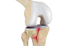
Knee Fracture
A fracture is a condition in which there is a break in the continuity of the bone. In younger individuals, these fractures are caused by high energy injuries, as from a motor vehicle accident. In older people, the most common cause is a weak and fragile bone.
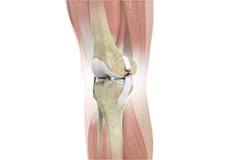
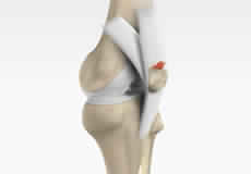
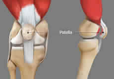
Patella Fracture
The kneecap or patella forms a part of the knee joint. It is present at the front of the knee, protecting the knee and providing attachment to various muscle groups of the thigh and leg. The undersurface of the kneecap and the lower end of the femur are coated with articular cartilage, which helps in smooth movement of the knee joint. A fracture in the kneecap is rare but common in adult males.
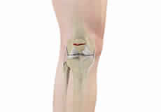
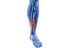
Tibial Shaft Fracture
A tibial shaft fracture is a crack or break in the middle section of the tibia bone due to severe trauma.
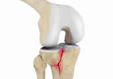
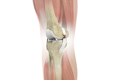
Tibial Plateau Fracture
A tibial plateau fracture is a crack or break on the top surface of the tibia or shinbone in the knee joint. The fracture most often occurs following a high-intensity trauma or injury from the impaction of the femoral condyles over the tibial plateau. Fractures of the tibial plateau are serious injuries that are more commonly seen in athletes involved in high-impact sports such as basketball, rugby, and football and can put athletes out of action for a long period of time.
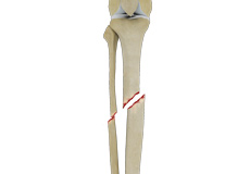
Fractures of the Tibia
The lower leg is made up of two long bones called the tibia and fibula that extend between the knee and ankle. The tibia or shinbone is the larger of the two bones. It bears most of the body’s weight and helps form the ankle joint and knee joint.
ACL Tears
The anterior cruciate ligament (ACL) is one of the major ligaments of the knee. It is located in the middle of the knee and runs from the femur (thighbone) to the tibia (shinbone). The ACL prevents the tibia from sliding out in front of the femur. Together with the posterior cruciate ligament (PCL), it provides rotational stability to the knee.
Knee Ligament Injuries
Knee problems may arise if any of these structures get injured by overuse or suddenly during sports activities. Pain, swelling, and stiffness are the common symptoms of any damage or injury to the knee.
Multiligament Instability
The knee is a complex joint of the body that is vital for movement. The four major ligaments of the knee are anterior cruciate ligament (ACL), posterior cruciate ligament (PCL), medial collateral ligament (MCL) and lateral collateral ligament (LCL). They play an important role in maintaining the stability of the knee. A multiligament injury is a tear in one or more ligaments of the knee, which affects the knee stability.
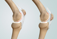
Patellar Dislocation/Patellofemoral Dislocation
Patellar dislocation occurs when the patella moves out of the patellofemoral groove, (trochlea) onto the bony head of the femur. If the kneecap partially comes out of the groove, it is called subluxation; if the kneecap completely comes out, it is called dislocation (luxation).
Patellar Instability
Any damage to the supporting ligaments may cause the patella to slip out of the groove either partially (subluxation) or completely (dislocation). This misalignment can damage the underlying soft structures such as muscles and ligaments that hold the kneecap in place. Once damaged, these soft structures are unable to keep the patella (kneecap) in position. Repeated subluxation or dislocation makes the knee unstable. This condition is called knee instability. Patellar (kneecap) instability results from one or more complete or partial dislocations (subluxations).
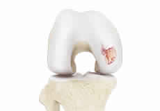
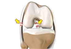
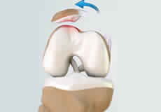
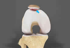
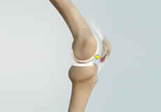
Chondral or Articular Cartilage Defects
The articular or hyaline cartilage is the tissue lining the surface of the two bones in the knee joint. Cartilage helps the bones move smoothly against each other and can withstand the weight of your body during activities such as running and jumping. Articular cartilage does not have a direct blood supply to it, so has less capacity to repair itself. Once the cartilage is torn it will not heal easily and can lead to degeneration of the articular surface, leading to the development of osteoarthritis.
Patellofemoral Instability
Patellofemoral instability means that the patella (kneecap) moves out of its normal pattern of alignment. This malalignment can damage the underlying soft structures such as muscles and ligaments that hold the knee in place.
Recurrent Patella Dislocation
The patella (kneecap) is a small bone that shields your knee joint. It is present in front of your knee, on a groove called the trochlear groove that sits at the junction of the femur (thighbone) and tibia (shinbone). Articular cartilage presents below the patella and end of the femur cushion and helps the bones glide smoothly over each other when the legs move. This joint is stabilized and supported by a network of soft tissues. The medial patellofemoral ligament (MPFL) connects to the inner side of the patella and helps to keep it from slipping away from the knee. Damage to this ligament leads to patellar dislocation.
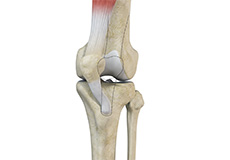
Quadriceps Tendon Rupture
The quadriceps can rupture after a fall, direct blow to the leg and when you land on your leg awkwardly from a jump. Quadriceps tendon rupture most commonly occurs in middle-aged people who participate in sports that involve jumping and running. Other causes include tendonitis (inflammation of quadriceps tendon), diseases such as rheumatoid arthritis, diabetes mellitus, infection and chronic renal failure, which weaken the quadriceps tendon. Use of medications such as steroids and some antibiotics also weakens the quadriceps tendon.
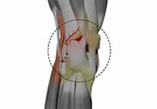
Posterolateral Instability
Posterolateral instability, also known as posterolateral rotatory instability (PLRI), is a common pattern of knee instability that results from injuries to the structures that support the outside of the knee joint, the posterolateral corner. When individuals injure the knee ligaments, they are most likely to injure the structures of the posterolateral corner. These structures include the popliteus tendon, lateral collateral ligament, and the knee joint capsule. The knee instability mostly occurs along with other ligamentous injuries, in particular, damage to either the ACL (anterior cruciate ligament), the PCL (posterior cruciate ligament), or both.
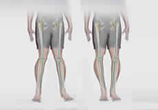
Knee Angular Deformities
Angular deformities of the knee are variations in the normal growth pattern during early childhood and are common during childhood.
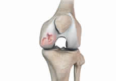
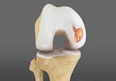
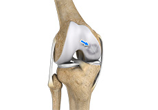
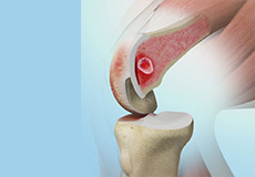
Osteochondral Defect of the Knee
An osteochondral defect, also commonly known as osteochondritis dissecans, of the knee refers to a damage or injury to the smooth articular cartilage surrounding the knee joint and the bone underneath the cartilage. The degree of damage may range from a rupture of the cartilage to a slight crack of the bone to a piece of the bone breaking off within the joint. These pieces may vary in size and depth and may remain stable or unstable within the joint. These may occur from an acute traumatic injury to the knee or an underlying disorder of the bone. Osteochondral defect is more common among young athletes who actively take part in sports and most commonly affects the femoral condyles in the knee.
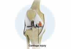
Articular Cartilage Injury
Articular or hyaline cartilage is the tissue lining the surface of the two bones in the knee joint. Cartilage helps the bones move smoothly against each other and can withstand the weight of the body during activities such as running and jumping. Articular cartilage does not have a direct blood supply to it so has little capacity to repair itself. Once the cartilage is torn it will not heal easily and can lead to degeneration of the articular surface, leading to the development of osteoarthritis.
Anterior Knee Pain
Anterior knee pain is characterized by chronic pain over the front and center of the knee joint. It is common in athletes, active adolescents (especially girls) and overweight individuals. Anterior knee pain refers to various conditions, which include runner's knee or patellar tendinitis, and chondromalacia of the patella. There is an inter-individual variation in the duration and presentation of pain.
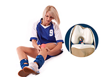
Chondromalacia Patella
Chondromalacia patella is a common condition characterized by softening, weakening and damage of the cartilage. The condition is most often seen in young athletes and older adults who have arthritis of the knee. It especially occurs in women.
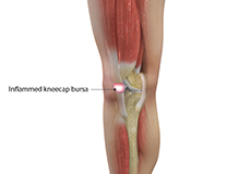
Kneecap Bursitis
Bursitis refers to the inflammation and swelling of the bursa. Inflammation of the bursa in front of the kneecap (patella) is known as kneecap bursitis or prepatellar bursitis.
Iliotibial Band Syndrome
Iliotibial band syndrome is an overuse injury resulting from the inflammation of the iliotibial band. It occurs when the iliotibial band and the lower outside portion of the thighbone at the knee joint rub against each other.
Lateral Patellar Compression Syndrome
Lateral patellar compression syndrome refers to pain under and around your kneecap. It is a common complaint among runners, jumpers and other athletes such as skiers, cyclists, and soccer players.
Osteochondritis Dissecans of the Knee
Osteochondritis dissecans is a joint condition in which a piece of cartilage, along with a thin layer of the bone separates from the end of the bone because of inadequate blood supply. The separated fragments are sometimes called “joint mice”. These fragments may be localized or may detach and fall into the joint space, causing pain and joint instability. Osteochondritis dissecans occurs within the lateral aspect of the medial femoral condyle. The condition can also occur in other joints, including your elbows, ankles, shoulders, and hips.
Runner's Knee
Patellofemoral pain syndrome also called runner’s knee refers to pain under and around your kneecap. Patellofemoral pain is associated with a number of medical conditions such as anterior knee pain syndrome, patellofemoral malalignment, and chondromalacia patella. Patellofemoral pain is a common complaint among runners, jumpers, and other athletes such as skiers, cyclists, and soccer players; thus the common name, runner’s knee.
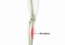
Shin Splints
Shin splints are pain and inflammation of the tendons, muscles and bone tissue along the tibia or shinbone (lower leg). It occurs because of vigorous physical activities such as exercise or sports. The condition is also referred to as medial tibial stress syndrome (MTSS).
MCL Sprains
MCL sprains occur due to a sudden impact from the outside of your knee, most commonly while playing sports such as rugby and football. Rarely, the MCL can get injured when the knee gets twisted or following a quick change in direction.
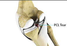
PCL Injuries
Posterior cruciate ligament (PCL), one of the four major ligaments of the knee, is situated at the back of the knee. It connects the thighbone (femur) to the shinbone (tibia). The PCL limits the backward motion of the shinbone.
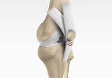
Patellar Tendon Rupture
The patellar tendon works together with the quadriceps muscle and the quadriceps tendon to allow your knee to straighten out. Patella tendon rupture is the rupture of the tendon that connects the patella (kneecap) to the top portion of the tibia (shinbone).
Lateral Meniscus Syndrome
Lateral meniscus syndrome is characterized by an injury caused by the tearing of the cartilage tissue or a rare case of a congenital abnormality called a discoid meniscus, which results in knee pain.
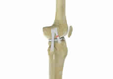
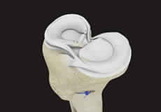
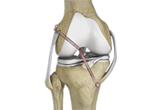
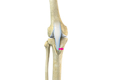
Tibial Eminence Spine Avulsion Fracture
Tibial eminence spine avulsion fracture is the avulsion (tearing away) of the tibial eminence.
Osteonecrosis of the Knee
Osteonecrosis is a condition in which the death of a section of bone occurs because of lack of blood supply to it. It is one of the most common causes of knee pain in older women. Women over 60 years of age are commonly affected, three times more often than men.
Knee Sports Injuries
Trauma is any injury caused during physical activity, motor vehicle accidents, electric shock, or other activities. Sports trauma or sports injuries refer to injuries caused while playing indoor or outdoor sports and exercising. Sports trauma can result from accidents, inadequate training, improper use of protective devices, or insufficient stretching or warm-up exercises. The most common sports injuries are sprains and strains, fractures, and dislocations.
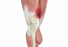
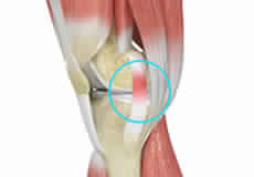
Patellar Tendinitis
Patellar tendinitis, also known as "jumper's knee", is an inflammation of the patellar tendon that connects your kneecap (patella) to your shinbone. This tendon helps in extension of the lower leg.
Women and ACL Injuries
The anterior cruciate ligament is one of the four major ligaments of the knee that connects the femur (thigh bone) to the tibia (shin bone) and helps stabilize the knee joint. Anterior cruciate ligament (ACL) injury is one of the common injuries of the knee. An injury to the ACL commonly occurs during sports or activities that involve twisting, overextension, landing from a jump incorrectly and abrupt change in direction or speed of movements.
Multiligament Knee Injuries
Injury to more than one knee ligament is called a multiligament knee injury and may occur during sports or other physical activities.
Osgood-Schlatter Disease
Osgood-Schlatter disease refers to a condition in older children and teenagers caused by excessive stress to the patellar tendon (located below the kneecap). Participants in sports such as soccer, gymnastics, basketball, and distance running are at higher risk for this disease.
Knee Arthroscopy
Knee arthroscopy is a common surgical procedure performed using an arthroscope, a viewing instrument, to diagnose or treat a knee problem. It is a relatively safe procedure and you will usually be discharged from the hospital on the same day of surgery.
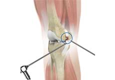
ACL Reconstruction
ACL (anterior cruciate ligament) reconstruction is a commonly performed surgical procedure. With recent advances in arthroscopic surgery, it can now be performed with minimal incision and low complication rates.
Posterolateral Corner (PLC) Reconstruction
A posterolateral corner is a complex arrangement of multiple ligaments, tendons, muscles and a joint capsule in your knee. It is located on the outside back corner of the knee.
Meniscus Replacement
Meniscus replacement is a surgical procedure performed to replace a torn or damaged meniscus in the knee. The meniscus is a soft, fibrous disk of cartilage in your knee joint that sits between the femur (thighbone) and the tibia (shinbone). Also called fibrocartilage, it cushions and stabilizes the joint, acting as a smooth surface for the joint to move on to prevent wear and tear. There are two menisci - one on each side of your knee joint.
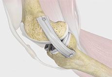
Knee Ligament Reconstruction
Knee ligament reconstruction is a surgical procedure to repair or replace damaged ligaments of the knee joint. The surgery can be performed using minimally invasive techniques.
Quadriceps Tendon Repair
Quadriceps tendon is a thick tissue located at the top of the kneecap. The quadriceps tendon works together with the quadriceps muscles to allow us to straighten our leg. The quadriceps muscles are the muscles located in front of the thigh.
Revision Knee Ligament Reconstruction
Revision knee ligament reconstruction is a complex surgical procedure performed to address failures or to correct the undesirable consequences of primary reconstruction surgery on your knee.
LPFL Reconstruction
LPFL reconstruction or lateral patellofemoral ligament reconstruction is a surgical procedure employed to treat patients with severe patellofemoral instability. The procedure involves replacing a torn lateral patellofemoral ligament with a part of a tendon taken from your leg. The main objective of the LPFL reconstruction is to tighten the knee joint and restore its stability.
Posterolateral Corner Reconstruction
Posterolateral corner injury is damage or injury to the structures of the posterolateral corner. The structures of the posterolateral corner include the lateral collateral ligament, the popliteus tendon, and the popliteo-fibular ligament. Injuries to the posterolateral corner most often occur with athletic trauma, motor-vehicle accidents, and falls. An isolated injury to the posterolateral corner is rare but often occurs with injuries to the cruciate ligaments, the anterior cruciate ligament (ACL) and the posterior cruciate ligament (PCL).
Multiligament Reconstruction of the Knee
Multiligament knee reconstruction is a surgical procedure to repair or replace two or more damaged ligaments of the knee joint. The surgery can be performed using minimally invasive techniques.
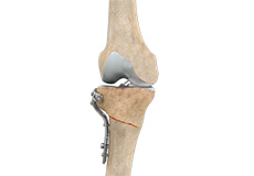
Knee Fracture Surgery
Knee fracture surgery is a surgical procedure performed to correct the cracked or broken bones in or around the knee to restore normal anatomical function, stability, and motion.
Meniscal Transplantation
Meniscal transplantation is a surgical procedure to replace the damaged meniscus of the knee with healthy cartilage. The meniscus is a C-shaped cartilage ring that acts as a cushion between the shinbone and the thighbone. Each of your knees has two menisci - one on the inside (medial aspect) and the other on the outside (lateral aspect)of your knee. Apart from the cushioning effect, the menisci also provide stability to the knee.
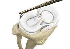
Meniscectomy
Meniscectomy is a surgical procedure indicated in individuals with torn meniscus where the conservative treatments are a failure to relieve the pain and other symptoms. Meniscectomy is recommended based on the ability of meniscus to heal, patient’s age, health status, and activity level.
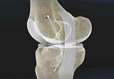
Quadriceps Tendon Autograft for Anterior Cruciate Ligament Reconstruction
Anterior cruciate ligament reconstruction using a quadriceps tendon autograft is a surgical procedure performed to replace a torn or damaged anterior cruciate ligament (ACL) of the knee with a part of the quadriceps tendon taken from your leg (autograft) to restore strength, stability, and function of the knee.
Partial Arthroscopic Meniscectomy
Partial arthroscopic meniscectomy is a procedure to remove the damaged part of a meniscus in the knee joint with the help an arthroscope. The meniscus is a C-shaped disc of cartilage between your thighbone and shinbone. There are 2 menisci in each knee. They act as shock absorbers and provide stability to the joint.
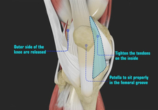
Patellofemoral Realignment
Patellofemoral realignment is a surgical procedure performed to treat symptomatic patellofemoral instability that does not respond to nonsurgical treatment measures.
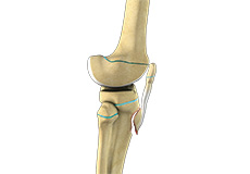
Tibial Tubercle Transfer
Tibial tubercle transfer is a surgical procedure that is performed along with other procedures to treat patellar instability, patellofemoral pain, and osteoarthritis. The tibial tubercle transfer technique involves realignment of the tibial tubercle (a bump in the front of the shinbone) such that the kneecap (patella) traverses in the center of the femoral groove. The patellar maltracking is corrected by moving the tibial tubercle medially, towards the inside of the leg. This removes the load off the painful portions of the kneecap and reduces pain.
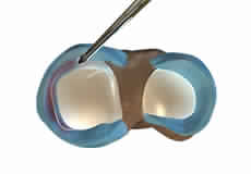
Lateral Lengthening
Lateral lengthening, also known as lateral retinacular lengthening or release, is a surgical procedure to release a tightened lateral retinaculum on the outer aspect of the knee. This procedure is mostly performed to treat knee pain or patellofemoral instability related to chronic pulling of the patella (kneecap) to the outer aspect of the knee, and the inability of the patella to rest properly in the center of the femoral groove as the knee bends and straightens.
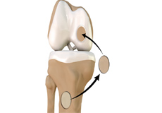
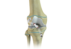
Mosaicplasty
Weight-bearing joints, such as the knee, may develop defects in the articular cartilage (spongy tissue that lines and cushions joints during movement) due to stress, trauma or degenerative disease. This can lead to pain, swelling or locking at the joint. Mosaicplasty is a surgical technique to repair the defect by transplanting healthy bone and cartilage from non-weight bearing areas of the knee. It is indicated to treat small cartilage defects of less than 2 cm in young active adults less than 45 years of age.
Arthroscopic Debridement
Arthroscopic debridement or a clean-up is a surgical procedure performed using an arthroscope. In this procedure, the cartilage or the bone that is damaged is removed using surgical instruments and the edges of the articular cartilage that are rough will be smoothened. A washout or joint lavage is done using a special tool to spray jets of fluid to wash and suck out to remove the remaining debris around the joint.
Distal Femoral Osteotomy
An osteotomy is a surgical procedure that involves cutting of bone. The distal femur is part of the femur (thighbone) just above the knee joint. Distal femoral osteotomy is performed to correct knee alignment which can lead to excessive loading and degeneration of one side of the knee joint. The procedure involves cutting of the distal femur, repositioning the bones and securing them in the proper alignment.
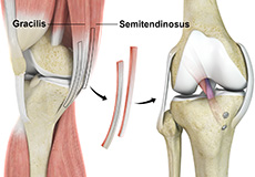
Hamstring Autograft
An autograft refers to using organ or tissue (bone, cartilage, tendon or skin) from the same person to transplant elsewhere on their body.
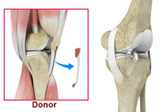
Hamstring Allograft
An allograft is an organ or tissue such as bone, cartilage, tendon or skin, taken from one person (donor) and surgically placed in another person to repair damaged tissue.
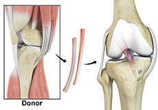
Bone-Patellar Tendon-Bone (BPTB) Allograft
ACL reconstruction with BPTP allograft is a surgical procedure that replaces the injured ACL with an allograft containing patellar tendon and bony attachments. The goal of the surgery is to tighten your knee and to restore its stability.

Physeal Sparing Reconstruction of the Anterior Cruciate Ligament
Physeal sparing reconstruction of the anterior cruciate ligament is a surgery to replace a torn anterior cruciate ligament or ACL, a major ligament of the knee, while minimizing damage to the growth plate (physis) present near the end of the bone. Ligaments are powerful bands of tissue that attach one bone to another, and the anterior cruciate ligament attaches the thighbone (femur) to the shinbone (tibia) to help stabilize the knee joint.
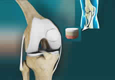
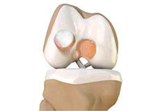
Partial Knee Resurfacing
Partial knee replacement is an alternative to total knee replacement in patients with arthritis on only one side of the knee. Partial knee replacement is a surgical procedure which involves resurfacing and replacement of only the diseased surface of the joint instead of the entire joint.
Prior Meniscectomy
The menisci are two C-shaped cartilages that act as shock absorbers between the thigh and shin bones that articulate at the knee joint. They provide stability and lubrication to the joint as well as nutrition for the articular cartilage. Tears in the meniscus may occur as a result of acute injury or chronic degeneration with age. Symptoms may include pain, swelling, and catching or locking of the joint. Meniscus tears can be surgically treated by meniscectomy.
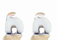
Trochleoplasty
Before performing trochleoplasty, your doctor will ensure that the articular cartilage (spongy tissue that lines and cushions joints during movement) at the trochlea is normal and healthy. The surgery is avoided if there is any sign of arthritis. The procedure may be performed as an open surgery or arthroscopically, a minimally invasive procedure that uses a narrow lighted tube with a camera to provide a clear view of the operating site. Trochleoplasty involves either lengthening the walls of the trochlear groove or deepening the groove by removing bone or any abnormal bony growths. You may sometimes need additional surgical procedures such as ligament reconstruction to improve the outcome.
Chondroplasty
Chondroplasty is a surgical procedure to repair and reshape damaged cartilage in a joint. The procedure involves smoothing degenerative cartilage and trimming any unstable flaps of cartilage.
Patellar Tendon Repair
Patellar tendon repair is the surgery performed to reattach the torn tendon to the kneecap and to restore normal function in the affected leg.
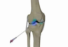
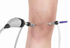
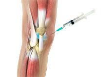
Intraarticular Knee Injection
Intra-articular knee injections are usually recommended when the pain has not responded to traditional conservative treatments such as anti-inflammatory medications, physical therapy, activity modification, or ice therapy.
Combined Hyaluronic Therapy for the Knee
Combined hyaluronic therapy is the process of injecting hyaluronic acid (HA) along with platelet-rich plasma (PRP) into your knee to treat osteoarthritis.
Knee Cartilage Restoration
Knee cartilage restoration is a surgical technique to repair damaged articular cartilage in the knee joint by stimulating new growth of cartilage or by transplanting cartilage into areas with defects in order to relieve pain and restore normal function to the knee.
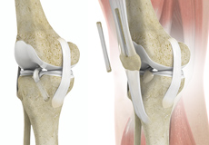
Knee Osteotomy
Knee osteotomy is a surgical procedure in which the upper shinbone (tibia) or lower thighbone (femur) is cut and realigned. It is usually performed in arthritic conditions affecting only one side of your knee. The aim is to take the pressure off the damaged area and shift it to the other side of your knee with healthy cartilage. During the surgery, your surgeon will remove or add a wedge of bone either below or above the knee joint, depending on the site of arthritic damage.
High Tibial Osteotomy
High tibial osteotomy is a surgical procedure performed to relieve pressure on the damaged site of an arthritic knee joint. It is usually performed in arthritic conditions affecting only one side of your knee and the aim is to take pressure off the damaged area and shift it to the other side of your knee with healthy cartilage. During the surgery, your surgeon will remove or add a wedge of bone either below or above the knee joint depending on the site of arthritic damage.
Tibial Tubercle Osteotomy
Tibial tubercle osteotomy is a surgical procedure that is performed along with other procedures to treat patellar instability, patellofemoral pain, and osteoarthritis. The tibial tubercle transfer technique involves realignment of the tibial tubercle (a bump in the front of the shinbone) such that the kneecap (patella) traverses in the center of the femoral groove. The patellar maltracking is corrected by moving the tibial tubercle medially, towards the inside of the leg. This removes the load off the painful portions of the kneecap and reduces pain.
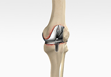
Unicompartmental/Partial Knee Replacement
Unicompartmental knee replacement is a minimally invasive surgery in which only the damaged compartment of the knee is replaced with an implant. It is also called a partial knee replacement.
What is New in Knee Replacement
Use of cementless parts that allow new bone to grow into a porous prosthesis and hold the parts in place, creating a biologic fixation
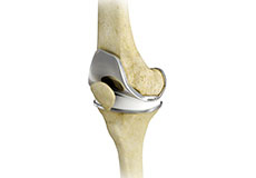
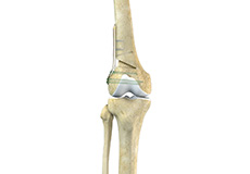
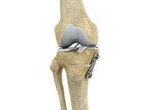
Total Knee Replacement
Total knee replacement, also called total knee arthroplasty, is a surgical procedure in which the worn out or damaged surfaces of the knee joint are removed and replaced with an artificial prosthesis.
Revision Knee Replacement
Revision knee replacement surgery involves replacing a part or all your previous knee prosthesis with a new prosthesis. Although total knee replacement surgery is successful, sometimes the procedure can fail due to various reasons and may require a second revision surgery.
Outpatient Total Knee Replacement
Total knee replacement is the surgical treatment for knee arthritis, where the damaged knee is removed and replaced with an artificial knee implant. Traditionally performed as an inpatient procedure, total knee replacement surgery is now being conducted on an outpatient basis, allowing you to go home on the same day of the surgery.
Robotic Assisted Partial Knee Surgery
Robotic-assisted partial knee surgery is an innovative alternative to the conventional surgical procedure to treat degenerative knee diseases such as osteoarthritis. It is performed using robotic-arm technology that allows your surgeon to precisely perform the surgery through small incisions.
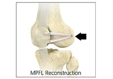
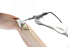
Medial Patellofemoral Ligament Reconstruction
Medial patellofemoral ligament reconstruction is a surgical procedure indicated for severe patellar instability.
Distal Realignment Procedures
Distal realignment procedures, also known as tibial tubercle transfer (TTT) procedures, are performed to reposition the kneecap after subluxation or dislocation by realigning the tendon under the kneecap to the underlying tibial tubercle.
Arthroscopic Reconstruction of the Knee for Ligament Injuries
Arthroscopic knee ligament reconstruction is a surgical procedure to correct a torn knee ligament by replacing the ligament with a healthy tendon tissue using an arthroscope.
PCL Reconstruction
PCL reconstruction surgery is a procedure to correct torn posterior cruciate ligament (PCL) in the knee using a tissue graft taken from another part of the body, or from a donor.
LCL Reconstruction
LCL reconstruction is a surgical procedure to repair torn or damaged lateral collateral ligament in the knee using a tissue graft taken from another part of the body, or from a donor.
MCL Reconstruction
MCL reconstruction is a minimally invasive surgical procedure in which a tendon graft is utilized to reconstruct the injured MCL.
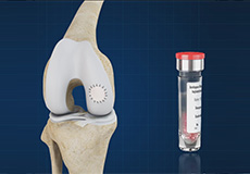
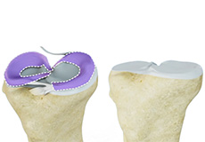
Autologous Chondrocyte Implantation
Autologous chondrocyte implantation (ACI) is a procedure to treat the articular cartilage defects of the knee. This procedure is effective for treating small areas of cartilage damage that causes pain and swelling and restricts range of motion. Autologous chondrocyte implantation is not indicated if you have advanced arthritis of the knee.
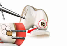
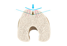
Subchondroplasty
Subchondroplasty is a minimally invasive procedure that is performed to specifically repair chronic BMLs by filling them with a bone substitute material. The bone substitute is then slowly resorbed and replaced with healthy bone, repairing the bone defect. Subchondroplasty also resolves the associated edema. Subchondroplasty may be performed alone or along with other arthroscopic procedures.
Partial Meniscectomy
Partial meniscectomy is a surgical procedure to remove the torn portion of the meniscus from the knee joint.
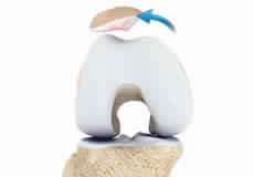
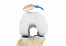
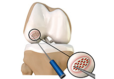
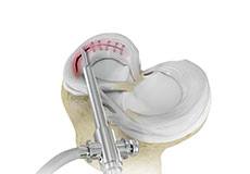
Cartilage Microfracture
Cartilage microfracture is a surgical procedure performed to replace the worn-out articular cartilage with new cartilage.
Meniscal Surgery
Meniscal surgery is a surgical procedure employed for the treatment of torn or damaged meniscal tissues in the knee. It is mostly performed as a minimally invasive keyhole procedure.
Partial Transphyseal Surgery
Partial transphyseal surgery is an arthroscopically assisted operative procedure to reconstruct the anterior cruciate ligament (ACL) in the knee of a child or adolescent with growth plates or physis (areas near the ends of long bones where growth is still occurring). Reconstruction of the ACL is performed using a soft tissue graft which is passed through tunnels drilled into the shinbone (tibia) and thighbone (femur) and secured to these bones.
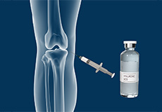
Viscosupplementation
Viscosupplementation refers to the injection of a hyaluronan preparation into the joint. Hyaluronan is a natural substance present in the joint fluid that assists in lubrication. It allows the smooth movement of the cartilage-covered articulating surfaces of the joint.
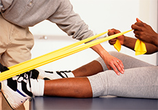
Physical Therapy for Knee
For knee problems, physical therapy involves strengthening and stretching certain joints and muscles. Physical therapy for the knee is designed to improve the muscle strength of the knee and reduce pain.

Pharmacological Interventions for Knee Injuries
Pharmacological interventions include medicinal preparations such as pain-relieving capsules or injections.
Non-Operative Treatments for ACL Injuries
Nonoperative treatments may be recommended if your child has a minor ACL injury, is still growing and does not wish to immediately return to sports.

Non-Surgical Knee Treatments
The knee is a complex joint which consists of bone, cartilage, ligaments, and tendons that make joint movements easy and at the same time it is more susceptible to various kinds of injuries. Knee problems may arise if any of these structures get injured by overuse or suddenly during sports activities. Injuries to the knee can be caused by degenerative diseases such as arthritis, traumatic injuries, and sports injuries. These conditions may affect the bones & joints and impair the mobility as well as the quality of life of the patients.
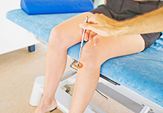
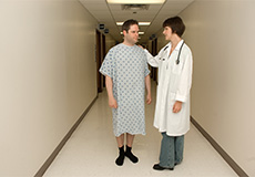
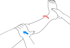
Physical Examination of the Knee
A complete physical examination of the knee is performed when you present to your doctor with a knee complaint. Both of your knees are examined and the results of the injured knee are compared to those of the healthy knee.
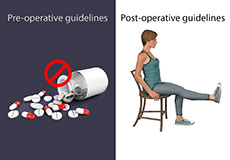
Pre-op and Post-op Knee Guidelines
Knee replacement is a surgery performed to replace parts of a diseased knee joint with an artificial prosthesis. The goal of knee replacement is to eliminate pain and return you to your normal activities. You can help in recovery and improve the outcomes of the procedure by following certain precautions and changing the way you carry out your daily activities.
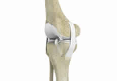
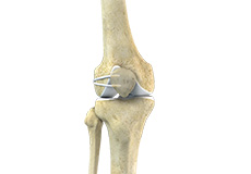
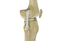
Knee Anatomy
The knee is a complex joint made up of different structures - bones, tendons, ligaments, and muscles. They all work together to maintain the knee’s normal function and provide stability to the knee during movement.
Having a well-functioning healthy knee is essential for our mobility and ability to participate in various activities. Understanding the anatomy of the knee enhances your ability to discuss and choose the right treatment procedure for knee problems with your doctor.
Bones of the Knee
The knee is a hinge joint made up of two bones, the thighbone (femur) and shinbone (tibia). There are two round knobs at the end of the femur called femoral condyles that articulate with the flat surface of the tibia called the tibial plateau. The tibial plateau on the inside of the leg is called the medial tibial plateau and on the outside of the leg, the lateral tibial plateau.
The two femoral condyles form a groove on the front (anterior) side of the knee called the patellofemoral groove. A small bone called the patella sits in this groove and forms the kneecap. It acts as a shield and protects the knee joint from direct trauma.
A fourth bone called the fibula is the other bone of the lower leg. This forms a small joint with the tibia. This joint has very little movement and is not considered a part of the main joint of the knee.
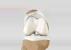
Articular Cartilage and Menisci of the Knee
Movement of the bones causes friction between the articulating surfaces. To reduce this friction, all articulating surfaces involved in the movement are covered with a white, shiny, slippery layer called articular cartilage. The articulating surface of the femoral condyles, tibial plateaus and the back of the patella are covered with this cartilage. The cartilage provides a smooth surface that facilitates easy movement.
To further reduce friction between the articulating surfaces of the bones, the knee joint is lined by a synovial membrane that produces a thick clear fluid called synovial fluid. This fluid lubricates and nourishes the cartilage and bones inside the joint capsule.
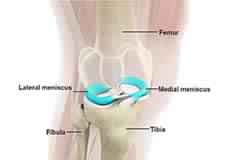
Within the knee joint, between the femur and tibia, are two C-shaped cartilaginous structures called menisci. Menisci function to provide stability to the knee by spreading the weight of the upper body across the whole surface of the tibial plateau. The menisci help in load-bearing i.e. it prevents the weight from concentrating onto a small area, which could damage the articular cartilage. The menisci also act as a cushion between the femur and tibia by absorbing the shock produced by activities such as walking, running and jumping.
Ligaments of the Knee
Ligaments are tough bands of tissue that connect one bone to another bone. The ligaments of the knee stabilize the knee joint. There are two important groups of ligaments that hold the bones of the knee joint together, collateral and cruciate ligaments.
Collateral ligaments are present on either side of the knee. They prevent the knee from moving too far during side to side motion. The collateral ligament on the inside is called the medial collateral ligament (MCL) and the collateral ligament on the outside is called the lateral collateral ligament (LCL).
Cruciate ligaments, present inside the knee joint, control the back-and-forth motion of the knee. The cruciate ligament in the front of the knee is called anterior cruciate ligament (ACL) and the cruciate ligament in the back of the knee is called posterior cruciate ligament (PCL).
Muscles of the Knee
There are two major muscles in the knee - the quadriceps and the hamstrings, which enable movement of the knee joint. The quadriceps muscles are located in front of the thigh. When the quadriceps muscles contract, the knee straightens. The hamstrings are located at the back of the thigh. When the hamstring muscles contract, the knee bends.
Tendons of the Knee
A tendon is a tissue that attaches a muscle to a bone. The quadriceps muscles of the knee meet just above the patella and attach to it through a tendon called the quadriceps tendon. The patella further attaches to the tibia through a tendon called the patella tendon. The quadriceps muscle, quadriceps tendon, and patellar tendon all work together to straighten the knee. Similarly, the hamstring muscles at the back of the leg are attached to the knee joint with the hamstring tendon.